Demonstration Model Of Medical Human Lumbar Intervertebral Disc, Intervertebral Disc Herniation Anatomical Model PVC Plastic Human Lumbar Disc Demonstration Model 11x10x7.6cm Medical Training Aid


Demonstration Model Of Medical Human Lumbar Intervertebral Disc, Intervertebral Disc Herniation Anatomical Model PVC Plastic Human Lumbar Disc Demonstration Model 11x10x7.6cm Medical Training Aid
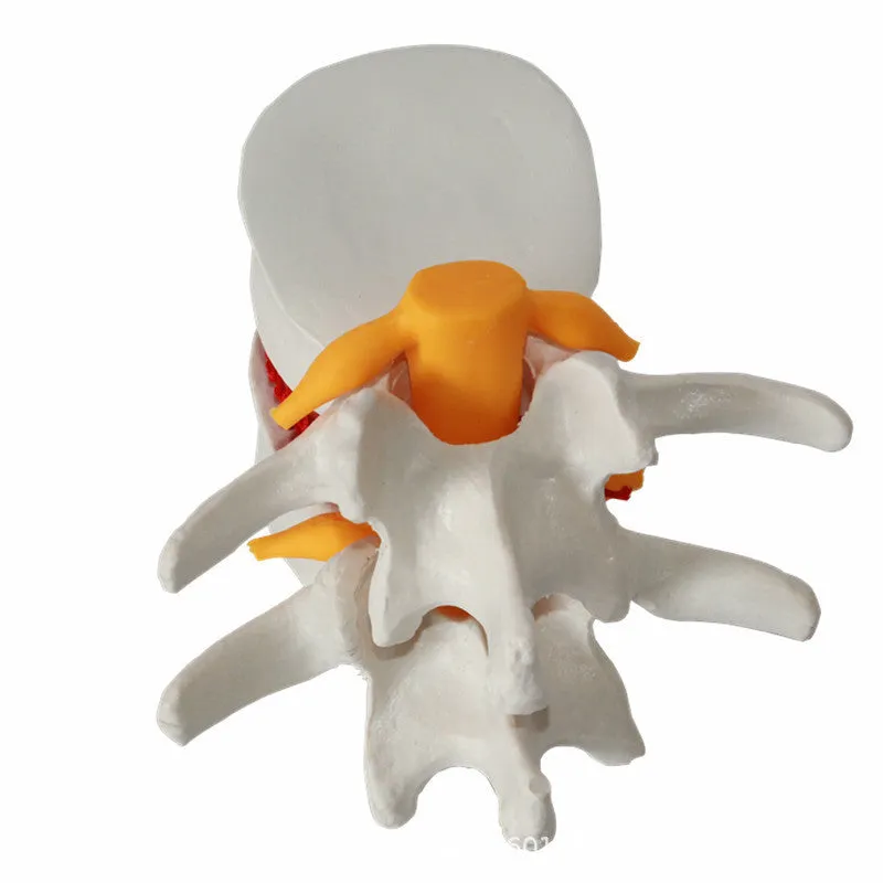
Medical Human Lumbar Intervertebral Disc Demonstration Model: 11x10x7.6cm PVC Anatomical Training Aid

This human lumbar intervertebral disc demonstration model serves as a focused anatomical reference tool specifically designed to illustrate intervertebral disc herniation conditions within the lumbar spine region. Measuring 11cm by 10cm by 7.6cm and constructed from moulded PVC plastic, it provides medical educators, students, and healthcare professionals with a tangible, three-dimensional visual aid that enhances understanding of lumbar disc pathology. The model's scale and detail support effective communication in clinical training environments, patient consultations, or educational settings where physical demonstration improves comprehension of spinal anatomy and common disc-related conditions. Its white finish ensures clear visibility of anatomical features under various lighting conditions, making it suitable for classroom presentations, clinic demonstrations, or individual study.
Features and Construction
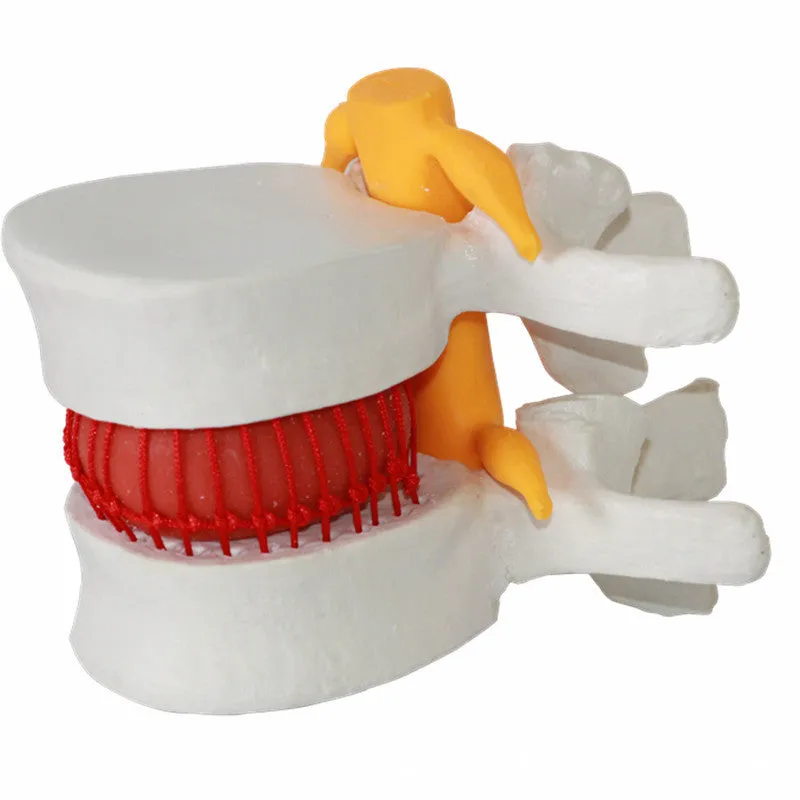
The demonstration model is engineered to provide a clear and accurate representation of lumbar intervertebral disc anatomy, with particular focus on herniation presentation. Its construction prioritises anatomical fidelity and practical handling for educational purposes, creating a reliable visual reference point for discussions about spinal health, disc pathology, and treatment approaches.
Material and Build
Constructed from PVC plastic, this model utilises material properties suited to anatomical demonstration tools. PVC allows for detailed moulding that captures the specific anatomical features of the lumbar intervertebral disc and surrounding structures relevant to herniation. The material offers sufficient durability for repeated handling in educational environments while maintaining the precise anatomical details necessary for accurate demonstration. This material choice supports the model's primary function as a visual and tactile aid during teaching sessions, consultations, or self-study without requiring specialist maintenance or care procedures.
Size and Practical Fit
With dimensions of 11cm in length, 10cm in width, and 7.6cm in height, the model presents a detailed yet manageable scale that facilitates clear observation of anatomical structures. The 0.22kg weight provides substantial handling feel without being cumbersome during demonstrations. This size-to-detail ratio makes the model suitable for individual examination, group presentations, or use alongside other anatomical references. The packaging dimensions of 11.5cm x 11cm x 8cm offer protective storage that preserves the model's anatomical details between uses, supporting its role as a reusable educational resource in various professional and academic settings.
Uses and Placement

This anatomical model serves specific functions within medical education and healthcare communication contexts. Its design supports both formal teaching environments and clinical explanation scenarios where visual aids enhance understanding of complex anatomical concepts.
Event or Professional Use
In professional healthcare settings, the model functions as a demonstration tool during patient consultations to visually explain lumbar disc herniation conditions, treatment options, or surgical procedures. Medical educators can incorporate it into anatomy lectures, physiotherapy training, or surgical preparation sessions where three-dimensional representation of disc pathology enhances learning outcomes. The model's specific focus on intervertebral disc herniation makes it particularly relevant for orthopaedic specialists, neurosurgeons, physiotherapists, and medical students studying spinal disorders, providing a concrete reference point that supplements textbook diagrams and imaging studies.
Everyday Home Use
While primarily designed for professional and educational environments, the model may also serve individuals pursuing anatomical study outside formal institutions. Medical students can use it for home revision of lumbar spine anatomy, healthcare professionals might employ it for personal reference, or patients with specific interest in their spinal conditions could utilise it to better understand medical explanations. Its self-contained presentation and clear anatomical detailing support focused study without requiring additional specialist equipment, making it accessible for various levels of anatomical interest when paired with appropriate educational materials.
Benefits and Buying Value

The model's value derives from its specific anatomical focus and practical construction, offering tangible benefits for those requiring visual demonstration tools in medical or educational contexts.
Reuse and Low Maintenance
The PVC construction and durable moulding support repeated use across multiple demonstration sessions, educational terms, or consultation appointments. Unlike perishable biological specimens or fragile replica materials, this model maintains its anatomical accuracy through handling, storage, and transportation between different usage environments. The simple material composition requires only basic care—careful handling and storage in the provided packaging—to preserve its demonstration utility over time, making it a cost-effective visual aid that avoids ongoing maintenance expenses or replacement costs associated with more delicate anatomical models.
Why Choose This Product
This demonstration model offers specific advantages for those requiring a focused anatomical reference for lumbar intervertebral disc herniation. Its 11x10x7.6cm dimensions provide optimal detail visibility within a manageable handling size, while the PVC material ensures anatomical fidelity without specialist preservation needs. The model addresses the practical requirement for three-dimensional visual aids in medical education and patient communication, filling a specific niche between simplified diagrams and complex full-spine models. For educators, clinicians, or students seeking a dedicated demonstration tool for disc pathology, this model delivers targeted anatomical representation supported by practical construction and clear visual presentation.